This set of Clinical Science Multiple Choice Questions & Answers (MCQs) focuses on “Echocardiography”.
1. Echocardiography is essentially ________
a) ultrasound of the heart
b) echoing sound of the heart
c) another name for a treadmill test
d) recording of heart sounds
View Answer
Explanation: The ultrasound of the heart is called the echocardiography. It provides a real time imaging of the heart with the help of the ultrasound. The various modes for imaging of the heart help detect various problems.
2. The probe used for imaging the heart is ________
a) Linear Probe
b) Curvilinear Probe
c) Phase Array Probe
d) Endocavitary Probe
View Answer
Explanation: This type of probe has 128 elements and each segment is used to transmit and receive data. Since every element can function individually, the user can change the shape and focal point of the ultrasonic beam. Their angle of inclination can also be changed. This allows the probe to image through the space of ribs and images the heart.
3. Which mode is used for cardiac imaging?
a) A – Mode
b) B – Mode
c) M – Mode
d) 3D – Mode
View Answer
Explanation: The M – Mode depicts the incoming image in a wave like a manner. This wave is generated by the moving parts of the body. Thus, the moving heart valve can be depicted in a wave like a manner and so any blockages or abnormalities can be detected with the irregular wave pattern.
4. Continuous Wave (CW), Pulsed Wave (PW) and Color Flow (CF) are terms used for which kind of ultrasound?
a) 3D ultrasound
b) Dynamic Ultrasound
c) Doppler Ultrasound
d) Advanced Ultrasound
View Answer
Explanation: The Doppler Ultrasound relies on the Doppler effect that happens with the sound waves to produce images. CW, PW and CF are the different kinds of ultrasounds that are projected in the body which result in different types of images.
5. Echocardiography can be used for even _________
a) remove embolus of the heart
b) correctly locate the blockages in the arteries and veins
c) assess the baby’s heart, anatomy and function
d) measure heartbeat
View Answer
Explanation: Ultrasound is a safe way that the doctors use to check the baby’s progress while in the womb. Echocardiography is the ultrasound of the heart and it can be done for an infant who is still in the mother’s womb. Fetal echocardiography is done to check the progress, growth and the health of the baby’s heart.
6. Which kind of echocardiography may be recommended to diagnose coronary heart disease?
a) Thoracic Echocardiography
b) Stress Echocardiography
c) Transesophageal Echocardiography
d) 3D Echocardiography
View Answer
Explanation: To check for a coronary heart disease, the person may get subjected to some sort of stress in the form of a physical exercise. The doctor will take an echocardiogram before the test and one after the test. For continuous monitoring, an ECG may be taken. Through this method, the strength of the heart muscles can be monitored.
7. Which of the following ultrasound techniques gives the best Ultrasound Image of the heart?
a) 2D phased array
b) Linear phased array
c) Transesophageal
d) Curvilinear array
View Answer
Explanation: This type of ultrasound is taken by inserting an endoscope with the transducer down the esophagus. The endoscope gets an unobstructed view of the heart through the esophagus and the ultrasound of the heart can be taken. It is difficult to take the ultrasound of the heart due to the ribs blocking the beams. To take the ultrasound through the chest, the acoustic window is too small and only specific angles can be taken. Transesophagal method is an invasive method to take the ultrasound of the heart.
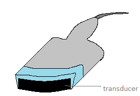
8. The following is an image of a curvilinear probe. What is best imaged with a curvilinear probe?
a) superficial structures and vessels
b) transabdominal imaging
c) cardiac imaging
d) transvaginal and transrectal imaging
View Answer
Explanation: The curve of the curvilinear probe increases the footprint i.e. the depth and surface area for imaging along with low frequency. Thus, it is used for taking the images of the abdomen. To take an image of the superficial structures and vessels, a linear probe is used as it can produce high frequency waves. Phase array probes are used for cardiac ultrasound as it can image between the ribs and endocavity probes are used for transrectal and transvaginal imaging.
Sanfoundry Global Education & Learning Series – Clinical Science.
To practice all areas of Clinical Science, here is complete set of 1000+ Multiple Choice Questions and Answers.
If you find a mistake in question / option / answer, kindly take a screenshot and email to [email protected]
- Check Clinical Science Books
- Practice Biotechnology MCQs
- Check Biotechnology Books
- Apply for Biotechnology Internship