This set of Clinical Science Multiple Choice Questions & Answers (MCQs) focuses on “Classification of Joints”.
1. The number of cartilagineous structures in a long bone are ____________
a) 2
b) 5
c) 0
d) 1
View Answer
Explanation: Each long bone has articular/hyaline cartilage at the outer extremities of the bone and an epiphysial cartilage at the extremities of the shaft. Hyaline Cartilage helps reduce friction and the epiphysial cartilage helps the bone grow. At maturity, the epiphysial cartilage of the bone solidifies.
2. The reason why people have joint pain in old age is because _____________
a) the bones become weak
b) the bones become too strong
c) the chondrocytes in articular cartilage become lesser in number
d) the blood supply to the joints reduces
View Answer
Explanation: Articular cartilage is the cartilage at the lining of the extremities of the long bones. It helps reduce friction and has chondrocytes. It has no blood supply and takes and nourishment from the synovial fluid. Drying of the synovial fluid and loss of chondrocytes causes pain in the joints.
3. Ellipsoid joint is found at __________
a) atlanto-occipital joint
b) first carpo-metacarple joint
c) first tarso-metatarsle joint
d) atlando-axial joint.
View Answer
Explanation: The elipsoid joint can perform functions like forward, backward and sliding movement. Atlanto-occipital joint is the joint between the 1st vertebra and the skull. Atlandto-axial joint is the joint of 1st and 2nd vertebrae. 1st carpo-metacarple joint and 1st tarso-metatarsle joints are the joints of the thumb and big toe.
4. Which of these is a multi-axial joint?
a) elbow joint
b) ball and Socket joint
c) shoulder joint
d) thumb joint
View Answer
Explanation: The term multiaxial joint stands for the joint which can move in all axis. Ball and socket joint can perform functions like an extension, flexion, abduction, aduction, rotation, circumduction, i.e., all possible movements. Thus, it’s multiaxial.
5. Other name for condylar joint is ___________
a) Saddle joint
b) Plain joint
c) Ball and Socket Joint
d) Hinge Joint
View Answer
Explanation: Condyle stands for a rounded articular surface on the extremity of bone where it forms a joint with another bone. This allows limited movements like extension and flexion. Since the movements are limited and possible in only one direction, i.e. uniaxial, it is also called a hinge joint.
6. In a synovial joint, the bones are connected to each other.
a) True
b) False
View Answer
Explanation: The bones are not connected to each other but held in place with the help of ligaments. A synovial capsule surrounds the joint and this capsule is filled with synovial fluid to reduce friction.
7. Synovial fluid secreted by synovial is rich in ___________
a) proteins
b) vitamins
c) minerals
d) fats
View Answer
Explanation: Synovial fluid is used to help reduce friction between the joints. Fats are good for cushioning and used as lining to avoid wear and tear. This helps to reduce friction. Thus, synovial fluid is rich in fats.
8. Acromioclavicular joint is an example of ___________
a) Ball and Socket Joint
b) Hinge Joint
c) Plain Joint
d) Pivot Joint
View Answer
Explanation: Acromioclavicular joint is the joint of the shoulder blades (scapula) and the shoulder bone (clavicle). This bone provides the sturdiness to the arm and thus can only perform the sliding action. Thus, the joint is called a plain joint.
9. The tooth like structure of C2 vertebra that forms Pivot Joint with C1 vertebra is called as odontoint. The other name is ____________
a) flagella
b) dens
c) matrix
d) condyl
View Answer
Explanation: The projecting tooth like structure is called as dens. Flagella is a whip like structure that allows single celled organism to move/swim. Condyl is a rounded structure at the extremity of the bones and matrix is a binding structure in which binds all the organic structures.
10. Label the diagram.
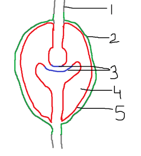
a) 1 – Periosteum, 2 – Fibrous Capsule, 3 – Hyaline Cartilage, 4 – Synovial Cavity, 5 – Synovial Capsule
b) 1 – Hyaline Cartilage, 2 – Fibrous Capsule, 3 – Synovial Cavity, 4 – Periosteum, 5 – Synovial Capsule
c) 1 – Hyaline Cartilage, 2 – Fibrous Capsule, 3 – Periosteum, 4 – Synovial Cavity, 5 – Synovial Capsule
d) 1 – Periosteum, 2 – Fibrous Capsule, 3 – Synovial Cavity, 4 – Hyaline Cartilage, 5 – Synovial Capsule
View Answer
Explanation: The following diagram is that of a freely moving joint or the synovial joint. The Synovial Joint is held together by ligaments. It has a synovial capsule and is surrounded by fiberous capsule. The synovial fluid is filled in the synovial capsule to help reduce friction. The hyaline cartilage at the moving parts also helps reduce wear and tear of the bones.
Sanfoundry Global Education & Learning Series – Clinical Science.
To practice all areas of Clinical Science, here is complete set of 1000+ Multiple Choice Questions and Answers.
If you find a mistake in question / option / answer, kindly take a screenshot and email to [email protected]
- Apply for Biotechnology Internship
- Check Biotechnology Books
- Check Clinical Science Books
- Practice Biotechnology MCQs